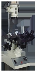
Product Description
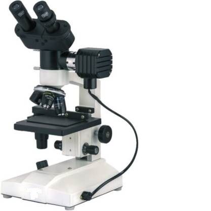
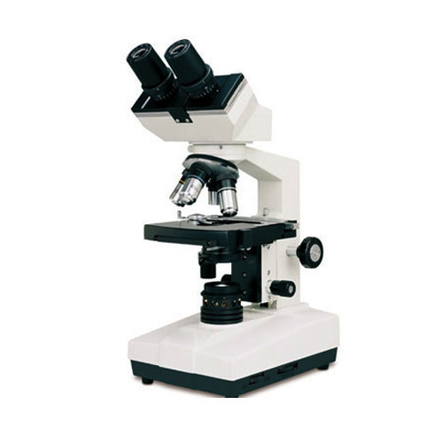
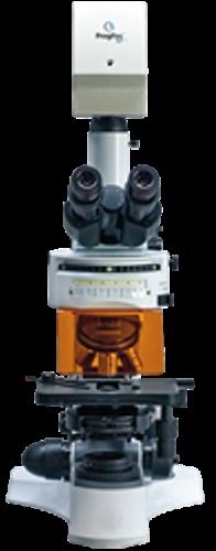
Inverted Tissue Culture Microscope with quadruple revolving nose piece, equipped with Trinocular Observation Head and best quality optical components having full prismatic optical path, large Co-axial Mechanical Stage of size 165mm. x 180mm., co-axial coarse & fine focussing knobs with adjustable tension control ring, variable intensity controlled built in base solid-state transformer, fitted with 6V 20Watt Halogen bulb and illumination based on Keohler's system.
Supplied with the following optical combination in a storing box.
- Observation Head - 45-degrees inclined Trinocular head interchangeable with Binocular Head.
- Nosepiece - Quadruple revolving turret nose piece with positive accurate click stops.
- Stage - With Large double plate coaxial mechanical stage.
- Illumination - Based on Koehler's system is provided by a post-mounted high intensity 6 volts 20 watts Halogen illuminator.
- Objectives - Achromatic 5x, 10x, LWD-20x & LWD-40XSL.
- Eyepieces - HWF 10x (paired) with eye guards & P7x for photo micrography.
Optimized Viewing for Tissue CultureSpecially designed for observation of living cells and tissues in culture dishes or flasks, this inverted microscope offers wide-field 10x eyepieces and achromatic objectives (10x, 20x, 40x) for sharp, high-resolution imaging. Its trinocular head, adjustable interpupillary distance, and comfortable inclined viewing make extended use fatigue-free.
Robust Construction and Ergonomic DesignBuilt with a heavy-duty die-cast metal body and anti-rust finish, the microscope ensures durability and stability in daily laboratory operations. Anti-fungal treated optics protect your investment, while smooth coaxial coarse and fine focus controls grant precise focusing every time.
Adaptable for Advanced Imaging and DocumentationWith a dedicated camera port, this microscope easily connects to digital cameras for effortless image and video documentation. Optional phase contrast kits and included color filters expand its versatility, making it a comprehensive tool for modern laboratory workflows.
FAQ's of Inverted Tissue Culture Microscope -C:
Q: How do I use the Inverted Tissue Culture Microscope-C for living specimen observation?
A: Simply place your culture dish or flask on the mechanical stage and use the coaxial coarse and fine focus knobs to bring your specimen into sharp focus. The inverted optical system allows easy access to the bottom of culture vessels, enabling clear views of living cells without disturbing the culture environment.
Q: What is the process for capturing digital images with this microscope?
A: Attach a compatible digital camera to the integrated camera port on the trinocular head. Once connected, you can use camera software (not included) to capture and document high-resolution still images or videos of your samples. The imaging quality depends on the attached camera model.
Q: When should I utilize the phase contrast kit, and how does it benefit observation?
A: The optional phase contrast kit is recommended when observing transparent or low-contrast specimens, such as unstained living cells. It enhances image contrast, revealing detailed structures that are otherwise difficult to distinguish under standard brightfield illumination.
Q: Where can I use this microscope, and what applications is it best suited for?
A: This microscope is ideal for laboratory settings, especially in cell biology, tissue culture, and research labs. Its ergonomic and stable design makes it suitable for routine analysis, observation of living cells, and documentation tasks requiring high optical resolution.
Q: What are the main benefits of the heavy-duty die-cast anti-rust body and anti-fungal optical treatment?
A: The anti-rust finish and die-cast metal construction ensure longevity and structural reliability even in humid laboratory environments. Anti-fungal treated optics safeguard the lenses and prisms, maintaining image clarity over long-term use.
Q: How do I adjust magnification and focus during operation?
A: Select your desired objective (10x, 20x, or 40x LWD achromatic) from the quadruple ball bearing nosepiece, then use the wide-field 10x eyepieces. Achieve precise focus using coarse and fine coaxial knobs, and adjust interpupillary distance and stage position for optimal viewing comfort and specimen access.


 English
 Spanish
 French
 German
 Italian
 Chinese (Simplified)
 Japanese
 Korean
 Arabic
 Portuguese